THE HEART
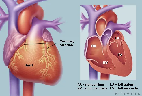
The heart is a muscular organ about the size of a fist, located just behind and slightly left of the breastbone. The heart pumps blood through the network of arteries and veins called the cardiovascular system.
The heart has four chambers:
The heart has four chambers:
- The right atrium receives blood from the veins and pumps it to the right ventricle.
- The right ventricle receives blood from the right atrium and pumps it to the lungs, where it is loaded with oxygen.
- The left atrium receives oxygenated blood from the lungs and pumps it to the left ventricle.
- The left ventricle (the strongest chamber) pumps oxygen-rich blood to the rest of the body. The left ventricle’s vigorous contractions create our blood pressure.
THE AORTA
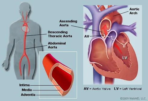
The aorta is the largest artery in the body. The aorta begins at the top of the left ventricle, the heart's muscular pumping chamber. The heart pumps blood from the left ventricle into the aorta through the aortic valve. Three leaflets on the aortic valve open and close with each heartbeat to allow one-way flow of blood.
The aorta is a tube about a foot long and just over an inch in diameter. The aorta is divided into four sections:
• The ascending aorta rises up from the heart and is about 2 inches long. The coronary arteries branch off the ascending aorta to supply the heart with blood.
• The aortic arch curves over the heart, giving rise to branches that bring blood to the head, neck, and arms.
• The descending thoracic aorta travels down through the chest. Its small branches supply blood to the ribs and some chest structures.
• The abdominal aorta begins at the diaphragm, splitting to become the paired iliac arteries in the lower abdomen. Most of the major organs receive blood from branches of the abdominal aorta.
Like all arteries, the aorta's wall has several layers:
• The intima, the innermost layer, provides a smooth surface for blood to flow across.
• The media, the middle layer with muscle and elastic fibers, allows the aorta to expand and contract with each heartbeat.
• The adventitia, the outer layer, provides additional support and structure to the aorta.
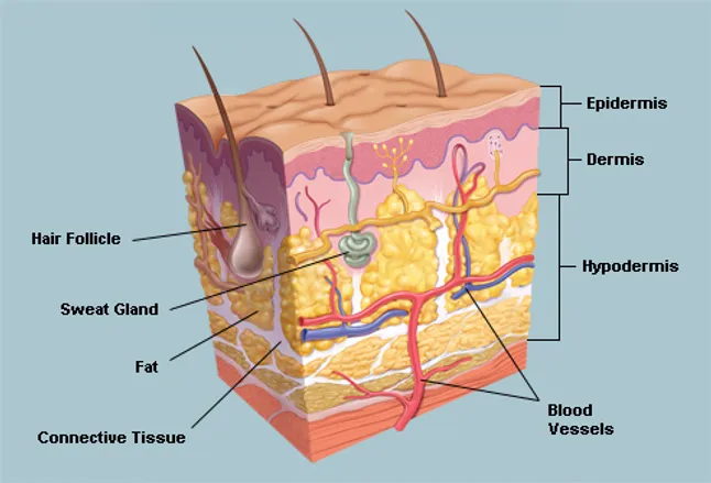
THE SKIN
The aorta is a tube about a foot long and just over an inch in diameter. The aorta is divided into four sections:
• The ascending aorta rises up from the heart and is about 2 inches long. The coronary arteries branch off the ascending aorta to supply the heart with blood.
• The aortic arch curves over the heart, giving rise to branches that bring blood to the head, neck, and arms.
• The descending thoracic aorta travels down through the chest. Its small branches supply blood to the ribs and some chest structures.
• The abdominal aorta begins at the diaphragm, splitting to become the paired iliac arteries in the lower abdomen. Most of the major organs receive blood from branches of the abdominal aorta.
Like all arteries, the aorta's wall has several layers:
• The intima, the innermost layer, provides a smooth surface for blood to flow across.
• The media, the middle layer with muscle and elastic fibers, allows the aorta to expand and contract with each heartbeat.
• The adventitia, the outer layer, provides additional support and structure to the aorta.
THE SKIN
The skin is the largest organ of the body, with a total area of about 20 square feet. The skin protects us from microbes and the elements, helps regulate body temperature, and permits the sensations of touch, heat, and cold.
Skin has three layers:
THE THYROID

Skin has three layers:
- The epidermis, the outermost layer of skin, provides a waterproof barrier and creates our skin tone.
- The dermis, beneath the epidermis, contains tough connective tissue, hair follicles, and sweat glands.
- The deeper subcutaneous tissue (hypodermis) is made of fat and connective tissue.
THE THYROID
The thyroid is a butterfly-shaped gland that sits low on the front of the neck. Your thyroid lies below your Adam’s apple, along the front of the windpipe. The thyroid has two side lobes, connected by a bridge (isthmus) in the middle. When the thyroid is its normal size, you can’t feel it.
Brownish-red in color, the thyroid is rich with blood vessels. Nerves important for voice quality also pass through the thyroid.
The thyroid secretes several hormones, collectively called thyroid hormones. The main hormone is thyroxine, also called T4. Thyroid hormones act throughout the body, influencing metabolism, growth and development, and body temperature. During infancy and childhood, adequate thyroid hormone is crucial for brain development.
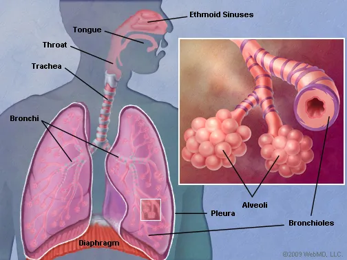
THE LUNGS
BRAIN
TRACHEA

Brownish-red in color, the thyroid is rich with blood vessels. Nerves important for voice quality also pass through the thyroid.
The thyroid secretes several hormones, collectively called thyroid hormones. The main hormone is thyroxine, also called T4. Thyroid hormones act throughout the body, influencing metabolism, growth and development, and body temperature. During infancy and childhood, adequate thyroid hormone is crucial for brain development.
THE LUNGS
The lungs are a pair of spongy, air-filled organs located on either side of the chest (thorax). The trachea (windpipe) conducts inhaled air into the lungs through its tubular branches, called bronchi. The bronchi then divide into smaller and smaller branches (bronchioles), finally becoming microscopic.
The bronchioles eventually end in clusters of microscopic air sacs called alveoli. In the alveoli, oxygen from the air is absorbed into the blood. Carbon dioxide, a waste product of metabolism, travels from the blood to the alveoli, where it can be exhaled. Between the alveoli is a thin layer of cells called the interstitium, which contains blood vessels and cells that help support the alveoli.
The lungs are covered by a thin tissue layer called the pleura. The same kind of thin tissue lines the inside of the chest cavity -- also called pleura. A thin layer of fluid acts as a lubricant allowing the lungs to slip smoothly as they expand and contract with each breath.
BRAIN
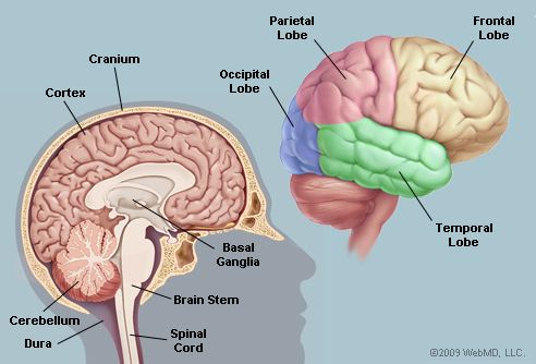
The brain is one of the largest and most complex organs in the human body.
It is made up of more than 100 billion nerves that communicate in trillions of connections called synapses.
The brain is made up of many specialized areas that work together:
• The cortex is the outermost layer of brain cells. Thinking and voluntary movements begin in the cortex.
• The brain stem is between the spinal cord and the rest of the brain. Basic functions like breathing and sleep are controlled here.
• The basal ganglia are a cluster of structures in the center of the brain. The basal ganglia coordinate messages between multiple other brain areas.
• The cerebellum is at the base and the back of the brain. The cerebellum is responsible for coordination and balance.
The brain is also divided into several lobes:
• The frontal lobes are responsible for problem solving and judgment and motor function.
• The parietal lobes manage sensation, handwriting, and body position.
• The temporal lobes are involved with memory and hearing.
• The occipital lobes contain the brain's visual processing system.
The brain is surrounded by a layer of tissue called the meninges. The skull (cranium) helps protect the brain from injury.
TRACHEA
The trachea, commonly known as the windpipe, is a tube about 4 inches long and less than an inch in diameter in most people. The trachea begins just under the larynx (voice box) and runs down behind the breastbone (sternum). The trachea then divides into two smaller tubes called bronchi: one bronchus for each lung.
The trachea is composed of about 20 rings of tough cartilage. The back part of each ring is made of muscle and connective tissue. Moist, smooth tissue called mucosa lines the inside of the trachea. The trachea widens and lengthens slightly with each breath in, returning to its resting size with each breath out.
The trachea is composed of about 20 rings of tough cartilage. The back part of each ring is made of muscle and connective tissue. Moist, smooth tissue called mucosa lines the inside of the trachea. The trachea widens and lengthens slightly with each breath in, returning to its resting size with each breath out.

0 comments